 Contacts
 Intranet
 SK
SK
The development of microtomography and its use in scientific research
Investigators: Miroslav Hain, Jozef Klembara
A modern 3D imaging method was developed – X-ray microtomography. We optimized the physical parameters of microtomographic measurements and subsequent digital data processing methods, which ensured the minimization and elimination of artifacts, noise reduction, increased image contrast, and effective image data segmentation. The development and use of this modern non-destructive measurement method in 2025 resulted in a publication in the scientific journal The Anatomical Record in the field of biology entitled: Comparative anatomy of the ossified braincase of legless anguine lizard Pseudopus apodus (Pallas, 1775). Based on data obtained from precise microtomographic measurements, this work described in detail the anatomy of the skull, inner ear, and individual bones of the braincase of the legless anguine Pseudopus apodus. Based on microtomographic data obtained from measurements of juvenile and adult individuals, the ontogenesis of its braincase was also described.
Projects: VEGA 1/0160/24, APVV-22-0328 (METIM), APVV-23-0366 (ARAM)
Publication:
- KLEMBARA, Jozef– HAIN, Miroslav. Comparative anatomy of the ossified braincase of legless anguine lizard Pseudopus apodus (Pallas, 1775) (Squamata, Anguimorpha). In The Anatomical Record, 2025. ISSN 1932-8486. (2.1 – IF2024) Q1
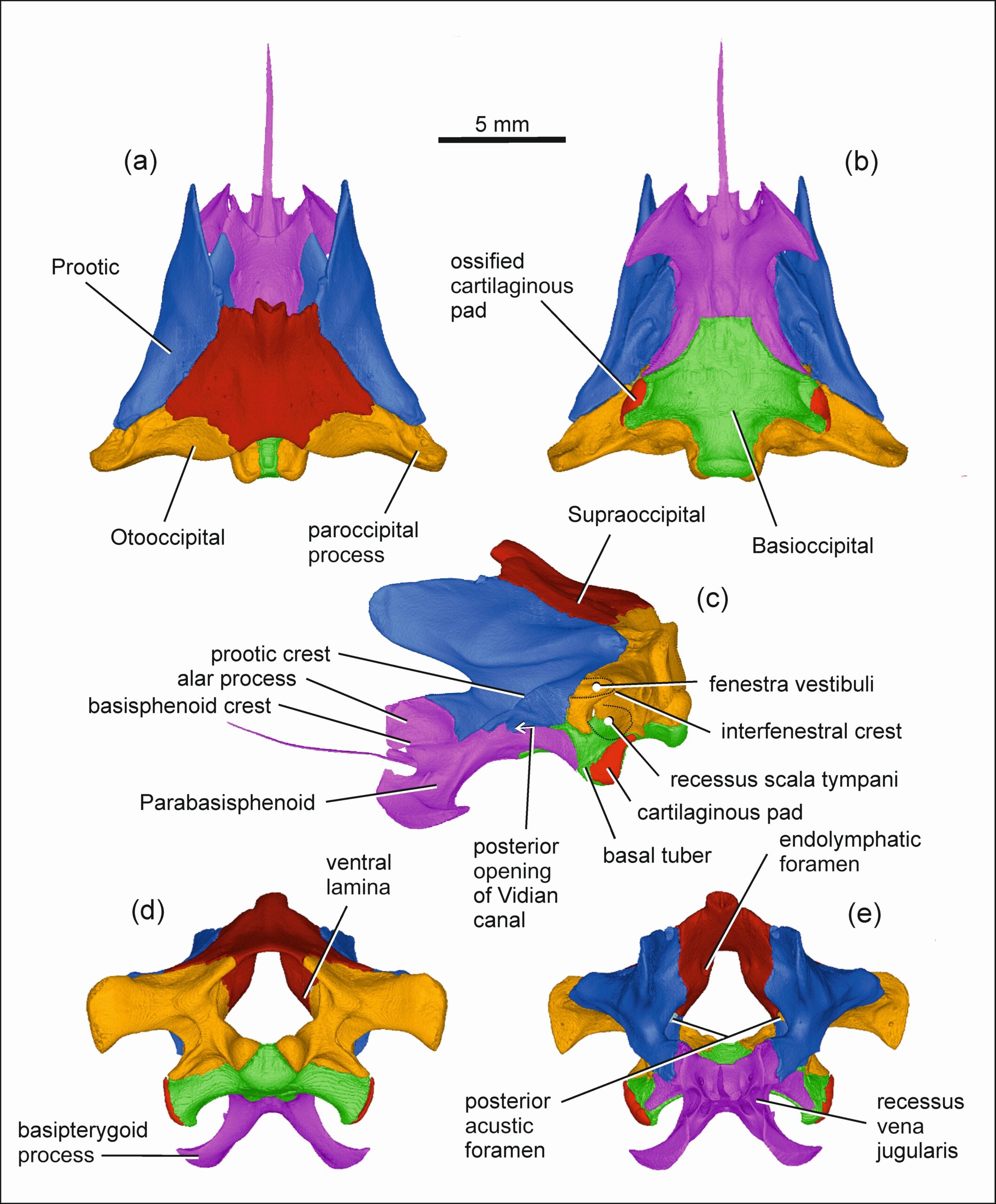
Fig.: 3D models of the ossified skull of Pseudopus apodus obtained by segmenting image data from microtomographic measurements