 Contacts
 Intranet
 SK
SK
X-ray microtomography in forensic science
The measurement and 3D imaging method of high-resolution X-ray microtomography – microCT developed at the SAV Measurement Institute has been successfully used in forensic science. In the close cooperation of ten scientific, university and criminological workplaces, the influence of the action of concentrated acid on the loss of dental mass, various selected dental fillings and bones was investigated using microCT, FTIR and other modern analytical methods with the use of artificial intelligence algorithms. An important part of the design of the measurement methodology was the optimization of the physical parameters of CT scans, especially in the case of amalgam fillings from the point of view of suppressing the appearance of reconstruction artifacts, which could significantly affect the resulting uncertainty of the measurements during the segmentation of the 3D image and the subsequent quantification of the segmented volumes. This research was primarily motivated by the investigation of a serious crime by the National Criminal Agency last year, when the Institute of Measurement with the microtomographic method of measurement was significantly involved in documenting this crime. The results of the work were published in the scientific journal Molecules (Q1), registered in Current Contents. Projects: COST CA 17121, COST CA 16101
Publication in CC magazine:
- THURZO, A. – JANČOVIČOVÁ, V. – HAIN, M. – THURZO, M. – NOVÁK, B. – KOSNÁČOVÁ, H. – LEHOTSKÁ, V. – VARGA, I. – KOVÁČ, P. – MORAVANSKÝ, N. Human remains identification using micro-CT, chemometric and AI methods in forensic experimental reconstruction of dental patterns after concentrated sulphuric acid significant impact. In Molecules, 2022, vol. 27, no. 13, p. 4035. ISSN 1420-3049. (2021: 4.927 – IF, Q2 – JCR, 0.705 – SJR, Q1 – SJR)
https://doi.org/10.3390/molecules27134035
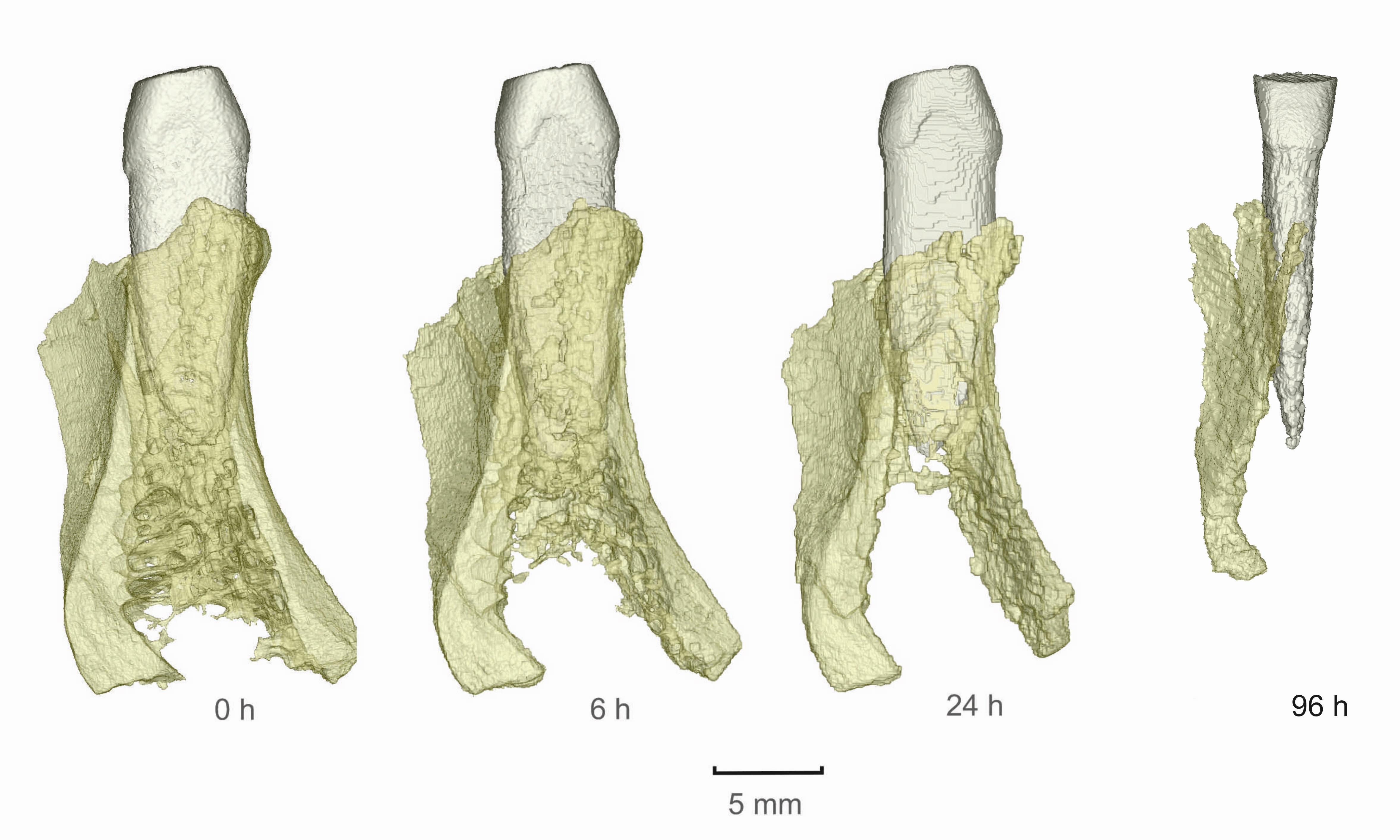
Fig.1: Time series of loss of tooth mass and bone after exposure to concentrated acid measured and displayed using X-ray microtomography (surface rendering, segmentation, bone displayed in semi-transparent mode)
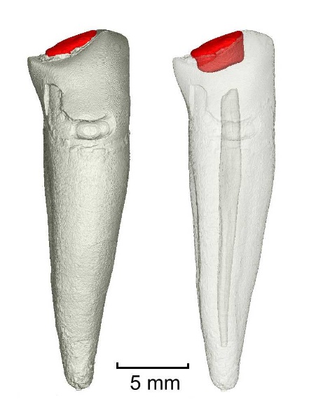
Fig.2: A tooth with a dental filling shown using X-ray microtomography (surface rendering, segmentation, transparent model on the right)