 Contacts
 Intranet
 SK
SK
Use of X-ray microtomography methods in comparative anatomy
Investigator: M. Hain
The X-ray microtomographic method of 3D imaging of biological objects was used in an extensive study focused on the comparative anatomy of legless lizards (Anguine Lizards) with emphasis on the species Pseudopus Apodus. Methods of data filtering, rendering and image segmentation were developed within the optimized microCT imaging.
The comparison showed significant morphological differences of Pseudopa vertebrates compared to Anguis and Ophisaurus, the results of which were published in CC journal [1].
Related projects:
• VEGA 1/0209/18 – Morphology of fossil lizards using computer microtomography imaging techniques, their phylogenetic relationships, paleobiogeography – migrations and community changes reflecting the gradual climatic changes of the Cenozoic.
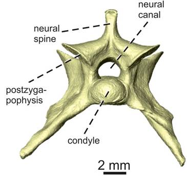
Fig. 1: Microtomographic image of the sacral vertebra Pseudopus Apodus
Result applicator:
• Faculty of Science, Charles University
Publications:
- ČERŇANSKÝ – YARYHIN O. – CICEKOVÁ J. – WERNEBURG I. – HAIN M. – KLEMBARA J.Vertebral Comparative Anatomy and Morphological Differences in Anguine Lizards With a Special Reference to Pseudopus apodus. In The anatomical record, 2018. DOI: 10.1002/ar.23944.