 Contacts
 Intranet
 SK
SK
Realistic simulation of heart activation and surface potentials while pacing the right ventricle
Investigators: Švehlíková, E. Cocherová, J. Zelinka, M. Haška, M. Tyšler
The activation of heart ventricles during right ventricular pacing was simulated in realistic patient heart geometry using activation models based on cellular automaton (CA) and reaction-diffusion equations (RD). Using the RD model, also normal ventricular activation was simulated that visually matched with the published experimental results in vivo. At the same time, different dynamics of the activation in both models could be observed. While constant velocity of activation front propagation was obtained in the CA model, in the RD model the velocity was dependent on the shape of the propagating wave front. Body surface potential maps during the first 20 ms of the simulated activation were computed in realistic model of the patient torso obtained from CT and compared with maps measured in the same patient during stimulation by a catheter placed in the right ventricle. The highest mean correlation between measured and simulated maps was 0.89 in homogeneous torso and 0.91 if the inhomogeneous torso model was used. These correlations were obtained when the position of the starting point of simulated activation was 23 and 22 mm from the real catheter position. This study showed, to what extent the CA and RD models can be used for simulation of the ventricular activation and verification of the results obtained when localizing the sources of ventricular arrhythmias.
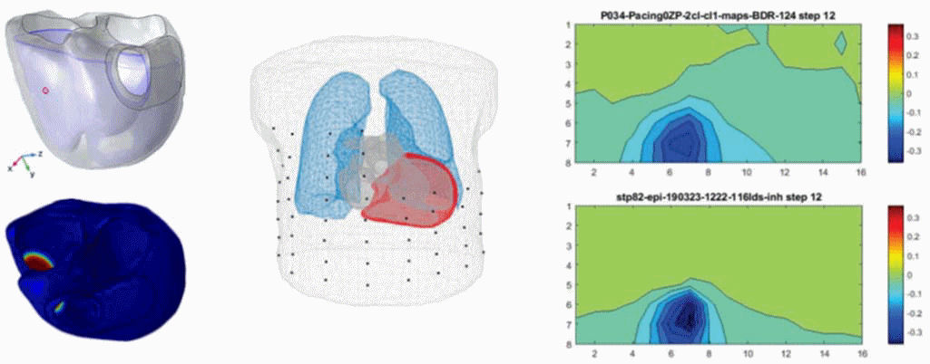
Figure: Left: Realistic heart geometry (frontal view, vertical heart position) with marked starting point of stimulated activation (top) and activated ventricular area 12 ms after stimulation (view from the heart base to apex). Center: inhomogeneous model of the patient torso and positions of measuring electrodes. Right: measured (top) and simulated map (bottom) of surface potentials [mV] 12 ms after the stimulation in the right ventricle.
Project:
VEGA 2/0125/19 – Measurement and modeling of the cardiac electrical field for noninvasive identification and interpretation of structural changes of the ventricular myocardium leading to ventricular arrhythmias.
Publications:
- ŠVEHLÍKOVÁ, Jana – ZELINKA, Ján – HAŠKA, Miroslav – TYŠLER Milan. Simulation of measured body surface potential map during early right ventricular pacing. In MEASUREMENT 2019 : Proceedings of the 12th International Conference on Measurement. Editors: J. Maňka, J. Švehlíková, V. Witkovský, I. Frollo. – Bratislava, Slovakia: Institute of Measurement Science, Slovak Academy of Sciences, 2019, p. 34-37. ISBN 978-80-972629-2-1.
- COCHEROVÁ, Elena – ŠVEHLÍKOVÁ, Jana – TYŠLER Milan. Evaluation of activation times in ventricular model with realistic geometry and conduction system. In MEASUREMENT 2019 : Proceedings of the 12th International Conference on Measurement. Editors: J. Maňka, J. Švehlíková, V. Witkovský, I. Frollo. – Bratislava, Slovakia: Institute of Measurement Science, Slovak Academy of Sciences, 2019, p. 26-29. ISBN 978-80-972629-2-1.
- COCHEROVÁ, Elena – ŠVEHLÍKOVÁ, Jana – TYŠLER, Milan. Activation in cardiac ventricular model with patient specific geometry and conduction system. In Trendy v biomedicínskom inžinierstve 2019 : 13. konferencia slovenských a českých pracovísk biomedicínskeho inžinierstva. – Žilina, Slovak Republic: University of Žilina, 2019. ISBN 978-80-554-1587-1.