 Department of Biomeasurements is oriented to research of measuring methods and devices for biology and medicine. Using models and computer simulations, methods for biosignals measurement and evaluation are being searched, possibility of their use for non-invasive assessment of the state and characteristics of selected biological objects are analyzed and special measuring devices are being developed enabling their application in biomedical research and in medical diagnostics and therapy.
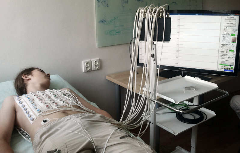
Current research is focused on multichannel measurement of cardiac electric field on the chest surface and is aimed for non-invasive assessment of physiological state of the heart and diagnostic and therapeutic exploitation of obtained information. Models of the heart and torso as an inhomogeneous volume conductor are beineg developed and processes of cardiac activation and generation of cardiac electric field in the torso are modelled. Possibilities of their use for forward ECG simulation and for inverse identification of selected pathologies are being verified, e.g. location of arrhythmogenic tissue or one or two local ischemic lesions in the myocardium. The research implies also evaluation of the sensitivity of developed solutions to errors in real measured data and to simplifications of used models.
Previous research activities of the department included analysis of gastric electrical activity, vascular pressures, signals from cardiovascular and neuromuscular system as indicators of thyroid gland function or measurement of electrograms and other physiological signals during experiments on isolated hearts of small animals.
Selected scientific results
-
A model for forward simulation of cardiac electric field was developed. The model enables definition of the ventricular myocardium with conduction system (in cooperation with Institute of Normal and Pathological Physiology SAS) and realistic geometry of inhomogeneous torso. Propagation of excitation in the myocardium is governed by cellular automaton or by eikonal equations (in cooperation with Università della Svizzera Italiana, Lugano, Switzerland). Potentials in the torso are computed using boundary element method and equivalent cardiac generator represented by homogeneous dipole layer or multiple dipole;

-
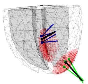
The ability of non-invasive identification of repolarization changes in the myocardium during local ischemia was analyzed using difference ECG integral maps a and one- or two-dipole model of the electrical source corresponding to the ischemic lesions. Possibilities of the method were tested using simulation experiments and verified on ischemic patients and patients that underwent cardiac intervention. The obtained results suggest that a mean lesion localization error of about 2 cm can be expected;

-
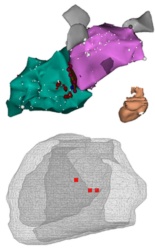
A method for noninvasive pre-operational localization of foci of premature ectopic ventricular activity using surface ECG mapping and CT or MR imaging was proposed. It is based on inverse solution with one equivalent dipole source. Dispersion of the found focus position is about 1 cm.
-
High resolution multi-channel measuring system for body surface potential mapping was developed. The new ProCardio 8 system uses active electrodes and can be used for non-invasive diagnostics of selected cardiac diseases based on potential and integral maps, including the above mentioned methods. The device is intended for biomedical research and for experimental clinical applications.
Cooperations
Department of Biomeasurements cooperates with medical and biomedical engineering research groups in Slovakia and several other countries. Currently they include namely:
- National Institute of Cardiovascular Diseases in Bratislava, Slovakia,
- Faculty of Biomedical Engineering CTU in Prague, Kladno, Czech Republic
- Institute of Biocybernetics and Biomedical Engineering PAS in Warszawa, Poland.
With several additional institutions and working groups there is an unformal cooperation. results of the coperation are common publications, exchange of developed programs, measured experimental results as well as common medical evaluation of proposed methods and measuring systems.
 Contacts
 Intranet
 SK
SK