 Contacts
 Intranet
 SK
SK
Method for accelerated simulation of electrical activity of anisotropic heart model using graphics processors
(Investigators: P. Kaľavský, M. Potse, S. Pezzuto, M. Tyšler)
A method for computationally fast simulation of electrical activation of an anisotropic model of the heart based on eikonal equations was proposed. The method was implemented in the parallel programming language CUDA and supports massive parallel computations on graphics processors (GPU). The computational speed of the proposed method was tested on four realistic heart models with a resolution of 1 mm, where it was able to simulate the activation sequence and a standard 12-lead ECG within 3 seconds. The simulated activation times and ECG signals were compared with values obtained from a reference reaction-diffusion model. The absolute error of the activation times in approximately 270,000 comparison points varied from 0 to 30 ms, but in 94% of the points it did not exceed 10 ms. When comparing simulated ECG signals, acceptable differences were obtained for QRS complexes, for which the average correlation coefficient was 85% and visually the vast majority of complexes matched in shape, polarity and duration and showed a difference of approximately 0.3 mV in the amplitude of the R-wave peak. Comparison of the T-waves of the ECG curves showed larger differences. The proposed method is actively used in the Institute of Computational Science, Universita della Svizzera Italiana in Lugano, Switzerland as part of inverse solutions for non-invasive characterization of ventricular activation abnormalities.
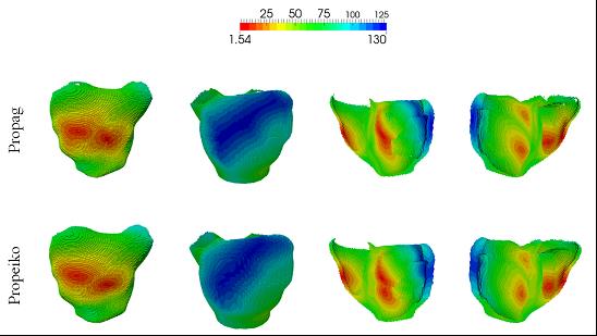
Fig. 1: Comparison of activation times in heart chambers obtained from the reference model (upper row) and using the proposed accelerated method based on eikonal equations (lower row). The images show a view of the heart from the front, from the back (left) and in a section from the front and from the back (right).
Related projects:
• VEGA 2/0071/16 “Modeling of the electric field of the heart to investigate the manifestations of functional and structural changes of the myocardium in the measured ECG signals.”
• APVV-14-0875 “Non-invasive localization of ectopic ventricular arrhythmias using ECG mapping and its use for causal treatment.”
• CATION – Cardiac activation time imaging for noninvasive characterization of ventricular conduction abnormalities, SCIEX-NMS project 14.158, Scientific Exchange Program between the New Members States of the EU and Switzerland.
Publication:
KAĽAVSKÝ Peter: GPU-accelerated model-based measuring methods for investigation of cardiac electrical activity. Dizertačná práca, Ústav merania SAV, 2016