 Contacts
 Intranet
 SK
SK
Modeling of possible factors influencing changes of QRS complex in ECG signals in left ventricular hypertrophy
(Investigators: J. Švehlíková, J. Zelinka, L. Bachárová, M. Tyšler)
Using a computer model of the ventricles, we simulated and visualized the spread of the activation front in the heart chambers at normal rate, in cases of local slowing of the activation queue, thickening of the left ventricular wall and a combination of both factors present in hypertrophy. The enlarged ECG signals of the QRS ventricular complex in hypertrophy are generally attributed to left ventricular enlargement. We have shown that the slowing of the activation of the activation front has a greater effect on signal amplification than the thickening of the left ventricular wall, which causes its longer presence in a large area of the myocardium, leading to an increase in potentials measured on the chest surface. The slowing of the excitation conduction or the thickening of the chamber wall by 50% did not cause a significant prolongation of the duration of the QRS complex. This occurred only when the spread of the activation front slowed down by 75% in a large part of the myocardium, resp. in combination with a slowing of the propagation propagation with a thickening of the left ventricular wall. This situation can occur with a combination of fibrosis and thickening of the heart wall of a hypertrophied heart. The obtained results point to the need to change the paradigm of the diagnosis of left ventricular hypertrophy.
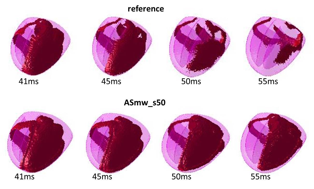
Fig. 1: Visualization of the activation queue at different time points of the QRS complex at normal propagation (top) and at a 50% deceleration of propagation in the anteroseptal region of the left ventricle (bottom).
Related projects:
• VEGA 2/0071/16 “Modeling of the electric field of the heart to investigate the manifestations of functional and structural changes of the myocardium in the measured ECG signals.”
• APVV-14-0875 “Non-invasive localization of ectopic ventricular arrhythmias using ECG mapping and its use for causal treatment.”
Publications:
- ŠVEHLÍKOVÁ, Jana – ZELINKA, Ján – BACHAROVA, L. – TYŠLER, Milan. Modeling and visualization of the activation wavefront propagation to improve understanding the QRS complex changes indicating left ventricular hypertrophy. In Journal of Electrocardiology, 2016, vol. 49, no. 5, p. 755-762. ISSN 0022-0736. (1.290-IF2015).
- BACHAROVA, L. – SZATHMARY, V. – ŠVEHLÍKOVÁ, Jana – MATEASIK, A. – GYHAGEN, J. – TYŠLER, Milan. The effect of conduction velocity slowing in left ventricular midwall on the QRS complex morphology: A simulation study. In Journal of Electrocardiology, 2016, vol. 49, no. 2, p. 164-170. ISSN 0022-0736. (1.290-IF2015).
- BACHAROVA, L. – SZATHMARY, V. – ŠVEHLÍKOVÁ, Jana – MATEASIK, A. – TYŠLER, Milan. QRS complex waveform indicators of ventricular activation slowing: Simulation studies. In Journal of Electrocardiology, 2016, vol. 49, no. 6, p. 790-793. ISSN 0022-0736. (1.290-IF2015).