 Contacts
 Intranet
 SK
SK
Biomedical research on the effects of the electromagnetic field at the cellular and subcellular level
Investigators: Michal Teplan, Hoang Vu Viet
In the area of the effect of pulsed electric fields (PEF) on sub-cellular structures, we investigated the manipulation of microtubules (MT) using PEF. In cooperation with ÚFE AVČR, we created new statistical measures for measuring MT movement for images from the “single molecule TIRF” microscope, which we called “microtubule displacement index” and “microtubule overlap rate” (see image). Using these measurements, we were able to show the effect of PEF voltage on MT displacement [1].
In the follow-up work [2], we focused on several key operations with a color image in the framework of preprocessing, segmentation and classification of microscopic images. We have also developed an innovative measurement approach to investigate MP effects on biological structures [3] based on continuous impedance spectroscopy. Potential uses of external electromagnetic fields include applications in diagnostics, therapy, and industry.
Foreign partner: Ing. Michal Cifra, PhD., Institute of Photonics and Electronics AS CR, Prague, Czech Republic
Related projects: Solved within the framework of projects MAD SAV-18-11, COST Action CA17115 and VEGA-2/0124/22.
Publications:
[1] HAVELKA, D. – ZHERNOV, I. – TEPLAN, Michal – LÁNSKÝ, Z. – CHAFAI, D.E. – CIFRA, M. Lab on chip microscope platform for electro manipulation of a dense microtubules network. In Scientific Reports, 2022, vol. 12, p. 2462. ISSN 2045-2322. (4.996 – IF2021) Q1
[2] BAJLA, Ivan – TEPLAN, Michal. Yeast cell detection in color microscopic images using ROC-optimized decoloring and segmentation. In IET Image Processing, 2022, vol. 16, no. 2, p. 606-621. ISSN 1751-9659. (1.773 – IF2021) Q2
[3] VU VIET, Hoang – TEPLAN, Michal. Impact of magnetic field on yeast cells monitored by impedance spectroscopy. In 2021 International Workshop on Impedance Spectroscopy (IWIS). – IEEE, 2022, p. 85-88. ISBN 978-1-6654-9472-4. Q4
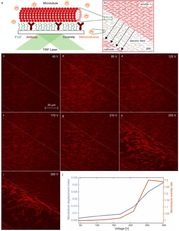
Figure: Dependence of microtubule displacement on pulse voltage. At each voltage, a series of 5 μs, N = 100 pulses with a frequency of 10 Hz was applied. a) MTs are bound to the coverslip surface via antibodies. (b) Schematic of the microscope field of view covering the electrode gap, (c)–(i) TIRF images show the MT (red) after μs-PEF treatment. Dashed lines represent the approximate position of the projection of the edges of the electrodes onto the image plane. (j) Microtubule displacement index and microtubule overlap rate. The data in this figure were collected sequentially on the same chip, sample, and field of view, starting at 45 V, then 85 V, 125 V, 170 V, 210 V, 255 V, and 300 V; thus, the observed effects were cumulative.