 Contacts
 Intranet
 SK
SK
New methods for determination of oxidative metabolism of skeletal muscle using phosphorus MR saturation transfer at rest and during exercise
(Investigators: L. Valkovič, I. Frollo)
Cardiovascular disease, obesity and diabetes are very often accompanied by changes in the energy metabolism of skeletal muscle. Therefore, modern medicine is placing increasing emphasis on research into new non-invasive methods for measuring energy metabolism. In line with this trend, new methodologies for determining oxidative metabolism using static and dynamic 31P-MR have been designed and validated. The utility of resting data measured on an MR system with a magnetic field intensity of 7 Tesla was demonstrated by comparing the metabolism of obese sedentary volunteers with trained individuals. The method is extremely topical for examinations of patients incapable of dynamic measurement (for example, after muscle injury). The proposed original methodology for measuring saturation transfer during a short (6 min.) Exercise brings a unique opportunity to determine the oxidative synthesis of ATP in muscle non-invasively. Such a measurement has great potential in research into human muscle metabolism and can lead to a significant improvement in the quality of life of an increasing number of patients.
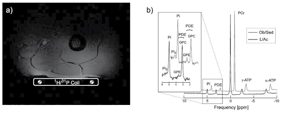
Fig. 1: Determination of innovative parameters of muscle energy metabolism. (a) a picture of the thigh muscle showing the position of the coil; (b) high-resolution 31P-MR spectra measured at rest in an obese sedentary (Ob / Sed) and trained (L / Ac) volunteer. The spectra are scaled to the intensity of the PCr signal and the resonance range of the Pi and PDE signals is approximated. Note the higher intensity of Pi2 and the lower intensity of PDE signals in the trained volunteer.
Related projects:
• VEGA 2/0013/14.
• APVV-0513-10, (Institute of Measurement SAS).
Publications:
- VALKOVIČ, Ladislav – CHMELÍK, M. – UKROPCOVÁ, B. – HECKMANN, T. – BOGNER, W. – FROLLO, Ivan – TSCHAN, H. – KREBS, M. – BACHL, N. – UKROPEC, J. – TRATTNIG S. – KRŠŠÁK, M. Skeletal muscle alkaline Pi pool is decreased in overweight-to-obese sedentary subjects and relates to mitochondrial capacity and phosphodiester content. In Scientific Reports, 2016, vol. 6, p. 20087. ISSN 2045-2322. (5.228-IF2015).
- VALKOVIČ, Ladislav – CHMELÍK, M. – MEYERSPEER, M. – GAGOSKI, B. – RODGERS, C.T. – KRŠŠÁK, M. – ANDRONESI, O.C. – TRATTNIG, S. – BOGNER, W. Dynamic 31P -MRSI using spiral spectroscopic imaging can map mitochondrial capacity in muscles of the human calf during plantar flexion exercise at 7 T. In NMR in Biomedicine, 2016, vol. 29, no. 12, p. 1825-1834. ISSN 0952-3480. (2.983 -IF2015).
- SCHMID, A.I. – MEYERSPEER, M. – ROBINSON, S.D. – GOLUCH, S. – WOLZT, M. – FIEDLER, G.B. – BOGNER, W. – LAISTLER, E. – KRŠŠÁK, M. – MOSER, E. – TRATTNIG, S. – VALKOVIČ, Ladislav. Dynamic PCr and pH imaging of human calf muscles during exercise and recovery using 31P gradient-Echo MRI at 7 Tesla. In Magnetic Resonance in Medicine, 2016, vol. 75, no. 6, p. 2324-2331. ISSN 0740-3194. (3.782-IF2015).