 Contacts
 Intranet
 SK
SK
Investigators: V. Juráš, P. Szomolányi, L. Valkovič
The goal of the research was to investigate T2 – relaxation parameters mapping as a possible marker for low-grade human articular cartilage lesions during a one-year follow-up, possible changes during the follow-up and compare the reliability and sensitivity of these measurements on high-field (3 T) and ultra-high-field (7 T) MRI scanners.
The change in T2 values acquired with 3 T using method “three-dimensional triple-echo steady-state” appears to be reflecting subtle changes of cartilage composition in the course of low-grade lesion development. 7 T T2 mapping does not reflect these changes probably due to completely decayed short T2 component.
The new method will find application in clinical practice and as a source for further research.
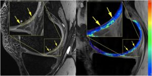
Fig. 1: An example of T2 maps and a cartilage bilayer segmentation of a patient with a low-grade cartilage lesion acquired at 7 T.
- A) Three-dimensional double-echo steady-state sequence image with the lesion delineated by the yellow arrows.
- B) T2 map overlaid on F0 contrast of a three-dimensional triple-echo steady-state image. The values of the color bars are T2 in milliseconds.
Related projects:
- ASC-2020, Comparative imaging methods based on magnetic resonance. Partner: Univ.-Prof. Dr. Siegfried Trattnig, MR Center, Highfield MR, Department of Radiology, Medical University of Vienna, Austria.
- VEGA 2/0001/17 – Measuring and imaging methods based on magnetic resonance for material and biomedical research
- APVV-15-0029 – Research of comparative imaging methods based on magnetic resonance for diagnostics of neurological and musculoskeletal diseases
Publications 2019:
- JURÁŠ, Vladimír – SCHREINER, M. – LAURENT, D. – ZBÝŇ, Š. – MLYNARIK, V. – SZOMOLÁNYI, Pavol – HAGER, B. – SCOTTI, C. – GOLDHAHN, J. – HEULE, R. – BIERI, O. – TRATTNIG, S. The comparison of the performance of 3 T and 7 T T2 mapping for untreated low-grade cartilage lesions. In Magnetic Resonance Imaging, 2019, vol. 55, p. 86-92. ISSN 0730-725X. (2.112-IF2018) Q1
- JURÁŠ, Vladimír – MLYNARIK, V. – SZOMOLÁNYI, Pavol – VALKOVIČ, Ladislav – TRATTNIG, S. Magnetic resonance imaging of the musculoskeletal system at 7T: Morphological imaging and beyond. In Topics in Magnetic Resonance Imaging, 2019, vol. 28, no. 3, p. 125-135. ISSN 0899-3459.
- HAGER, B. – WALZER, S.M. – DELIGIANNI, X. – BIERI, O. – BERG, A. – SCHREINER, M.M. – ZALAUDEK, M. – WINDHAGER, R. – TRATTNIG, S. – JURÁŠ, Vladimír. Orientation dependence and decay characteristics of T2* relaxation in the human meniscus studied with 7 Tesla MR microscopy and compared to histology. In Magnetic Resonance in Medicine, 2019, vol. 81, no. 2, p. 921-933. ISSN 0740-3194. (3.858-IF2018) Q1
- SZOMOLÁNYI, Pavol – ROHRER, M. – FRENZEL, T. – NOEBAUER-HUHMANN, I.M. – JOST, G. – ENDRIKAT, J. – TRATTNIG, S. – PIETSCH, H. Comparison of the relaxivities of macrocyclic gadolinium-based contrast agents in human plasma at 1.5, 3, and 7 T, and blood at 3 T. In Investigative Radiology, 2019, vol. 54, no. 9, p. 559-564. ISSN 0020-9996. (6.091-IF2018) Q1
- BRISTELA, M. – SKOLKA, A. – EDER, J. – SZOMOLÁNYI, Pavol – WEBER, M. – PIEHSLINGER, E. – SCHMID-SCHWAP, M. – TRATTNIG, S. T2 mapping with 3.0 T MRI of the temporomandibular joint disc of patients with disc dislocation. In Magnetic Resonance Imaging, 2019, vol. 58, p. 125-134. ISSN 0730-725X. (2.112-IF2018) Q1
- RAUDNER, M, – SCHREINER, M. M., – JURAS, V. –WEBER, M. – STELZENEDER, D. –KRONNERWETTER, C. –WINDHAGER, R. –TRATTNIG, S. Prediction of Lumbar Disk Herniation and Clinical Outcome Using Quantitative Magnetic Resonance Imaging. A 5-Year Follow-Up Study. Investigative Radiology • Volume 54, Number 3, March 2019. p. 183-189. (6.091-IF2018) Q1