 Contacts
 Intranet
 SK
SK
New methods of ensuring homogeneous signal excitation for determination of energy metabolism of skeletal muscles, liver and heart on 7T MRI tomography
(Investigators: L. Valkovič, I. Frollo)
Phosphorus MR spectroscopy (31P-MRS) is a unique technique that allows non-invasive measurement of tissue energy metabolism in vivo. However, due to the low signal-to-noise ratio (SNR), it is still not commonly used in medical practice. MR systems with a magnetic field strength of 7T in combination with surface coils achieve sufficient SNR, but lead to very inhomogeneous excitation, which makes data quantification considerably difficult. Therefore, new methodologies for ensuring homogeneous signal excitation have been designed and validated.
First of all, a new excitation pulse capable of homogeneous excitation despite an inhomogeneous field was designed. This new pulse was implemented in the measurement sequence and used to quantify cardiac metabolism in healthy volunteers. High reproducibility of the results was demonstrated even at different excitation field intensities.
A new technology providing a homogeneous excitation field for 31P-MRS of the whole body at 7T was also proposed. The performed magnetic field simulations were confirmed by measurements on healthy volunteers, where sufficient SNR was achieved by combining whole-body excitation with local signal uptake. This technology has great potential in research on human cardiac metabolism and can lead to an increase in the quality of life of many patients.
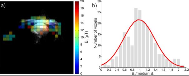
Fig. 1. In vivo excitation field mapping. (a) a representative map of the excitation field through the human body obtained using new technology. Sufficient SNR is only close to the receiving coil but shows high homogeneity. (b) histogram of the variability of the measured intensity of the excitation magnetic field with a normal distribution curve.
Foreign partner:
• Univ.-Prof. Dr. Siegfried Trattnig, MR Center, Highfield MR, Department of Radiology, Medical University of Vienna, Austria. Agreement on scientific cooperation until 31. XII. 2020th
Related projects:
• APVV-15-0029: “Research of comparative imaging methods based on magnetic resonance for the diagnosis of neurological and musculoskeletal diseases”.
• VEGA 2/0001/17: Measurement and imaging methods based on magnetic resonance for materials and biomedical research.
Publications:
- VALKOVIČ, L. – DRAGONU, I. – ALMUJAYYAZ, S. – BATZAKIS, A. – YOUNG, L.A.J. – PURVIS, L.A.B. – CLARKE, W.T. – WICHMANN, T. – LANZ, T. – FROLLO, I. – NEUBAUER, S. – ROBSON, M.D. – KLOMP, D.W.J. – RODGERS, C.T. Phosphorus Spectroscopic Imaging and B1+ mapping of human heart using a whole body transmit coil with 16-channel receive array at 7T. In Magnetic Resonance Materials in Physics, Biology and Medicine, 2017, vol. 30, suppl 1, p.S248-249. ISSN 1352-8661 (1.718-IF2016).
- VALKOVIČ, L. – CLARKE, W.T. – PURVIS, L.A.B. – SCHALLER, B. – ROBSON, M.D. – RODGERS, C.T. Adiabatic excitation for 31P MR spectroscopy in the human heart at 7 T: A feasibility study. In Magnetic Resonance in Medicine, 2017, vol. 78, no. 5, p. 1667-1673. ISSN 0740-3194. (3.924-IF2016)
- VALKOVIČ, L. – CHMELÍK, M. – KRŠŠÁK, M. In-vivo 31P-MRS of skeletal muscle and liver: A way for non-invasive assessment of their metabolism. In Analytical Biochemistry, 2017, vol. 529, p. 193-215. ISSN 0003-2697. (2.334-IF2016).
- VALKOVIČ, L. – DRAGONU, I. – ALMUJAYYAZ, S. – BATZAKIS, A. – YOUNG, L.A.J. – PURVIS, L.A.B. – CLARKE, W.T. – WICHMANN, T. – LANZ, T. – NEUBAUER, S. – ROBSON, M.D. – KLOMP, D.W.J. – RODGERS, C.T. Using a whole-body 31P birdcage transmit coil and 16-element receive array for human cardiac metabolic imaging at 7T. In PLoS ONE, 2017, vol. 12, no. 10, e0187153. ISSN 1932-6203. (2.806-IF2016).