 Contacts
 Intranet
 SK
SK
Modern investigation of cartilage damage using quantitative magnetic resonance
(Investigators: V. Juráš, P. Szomolányi, I. Frollo)
Although magnetic resonance imaging is currently considered the most accurate method for investigating cartilage damage, morphological imaging is often not sufficient to determine the degree of damage, nor does it detect early stages of degeneration, which are manifested solely by changes in the internal structure of cartilage. Quantitative MRI provides methods by which the state of collagen (T2 mapping) and proteoglycans (sodium MRI) can be analyzed. The result of the solution was a new method for mapping the relaxation constant T2 (based on the so-called steady-state free precision), which allows acceleration of the measurement (shortening from about 4 minutes to 2 minutes compared to the conventional method), higher resolution (factor 1.5) and an increase in the number of slices (which is most evident in ultra-high fields, 32 can be displayed instead of 4 slices). This method was used to detect differences in the collagen matrix in the cartilage of the knee and ankle, which has a wide application in clinical practice in the examination of cartilage (Fig. 1). Another modern method was used to determine proteoglycans – sodium MR, which instead of protons displays sodium ions that are directly proportional to the amount of proteoglycans. Since the reduction of proteoglycans in cartilage is the first sign of the onset of cartilage degeneration, this method has great potential to become part of screening measurements in clinical practice.
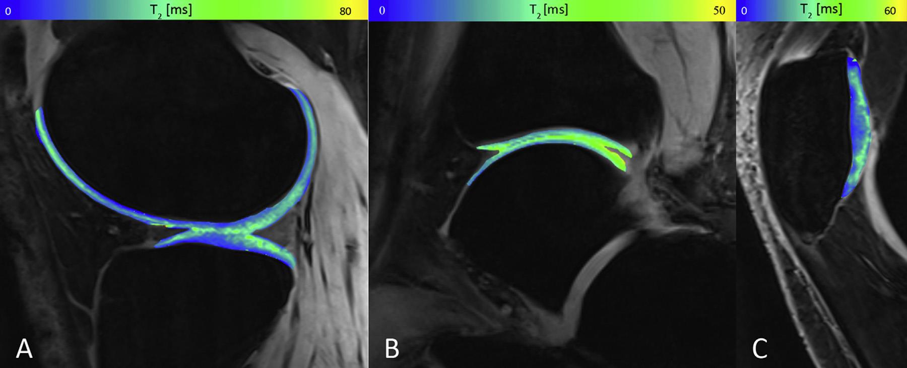
Fig. 1: Collagen fiber distribution and water distribution in cartilage measured by T2 mapping: A) femoral and tibial cartilage, B) ankle cartilage, C) patellar cartilage.
Related project:
• APVV 0431-12. (Institute of Measurement SAS).
Foreign partner:
• Univ.-Prof. Dr. Siegfried Trattnig, MR Center, Highfield MR, Department of Radiology, Medical University of Vienna, Austria.
Publications:
- JURÁŠ, Vladimír – ZBÝŇ, Š. – MLYNÁRIK, V. – SZOMOLÁNYI, Pavol – HAGER, B. – BAER, P. – FROLLO, Ivan – TRATTNIG, S. The compositional difference between ankle and knee cartilage demonstrated by T2 mapping at 7 Tesla MR. In European Journal of Radiology, 2016, vol. 85, no. 4, p. 771-777. ISSN 0720-048X. (2.593-IF2015)
- JURÁŠ, Vladimír – BOHNDORF, K. – HEULE, R. – KRONNERWETTER, C. – SZOMOLÁNYI, Pavol – HAGER, B. – BIERI, O. – ZBÝŇ, Š. – TRATTNIG, S. A comparison of multi-echo spin-echo and triple-echo steady-state T2 mapping for in vivo evaluation of articular cartilage. In European Radiology, 2016, vol. 26, no. 6, p. 1905-1912. ISSN 0938-7994. (3.640-IF2015)
- ZBÝŇ, Š. – MLYNÁRIK, V. – JURÁŠ, Vladimír – SZOMOLÁNYI, Pavol – TRATTNIG, S. Evaluation of cartilage repair and osteoarthritis with sodium MRI. In NMR in Biomedicine, 2016, vol. 29, no. 2, p. 206-215. ISSN 0952-3480. (2.983-IF2015)